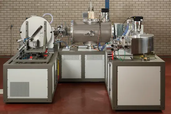
Multi-omics Mass Spectrometry Facility
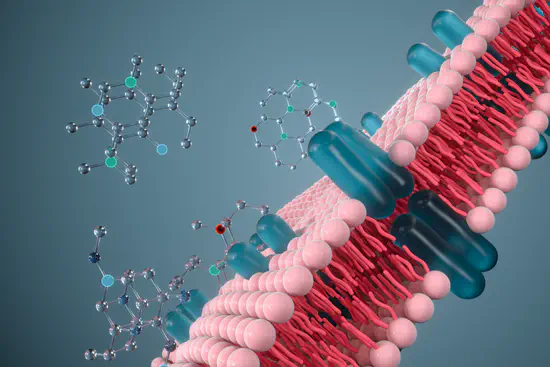
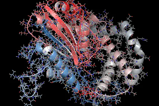
Our facility is equipped with the latest mass spectrometry technologies and staffed by a team of experts dedicated to advancing the understanding of complex biological systems. We integrate multiple omics strategies, from metabolomics to lipidomics and proteomics, culminating in the transformative approach of imaging mass spectrometry.